Home » Uncategories » Leg Muscle Diagram / HanhChampion Blogspot: Basic Leg Exercises / The gastrocnemius is the larger calf muscle, forming the bulge visible beneath the skin.
Leg Muscle Diagram / HanhChampion Blogspot: Basic Leg Exercises / The gastrocnemius is the larger calf muscle, forming the bulge visible beneath the skin.
Leg Muscle Diagram / HanhChampion Blogspot: Basic Leg Exercises / The gastrocnemius is the larger calf muscle, forming the bulge visible beneath the skin.. This chart is beautifully illustrated and offers the most comprehensive look at the muscles of the human leg available. It is also visible on the medial edge of the thigh from the anterior. Posterior muscles, such as the hamstrings and gluteus maximus, produce the opposite motion — extension of the thigh at the hip and flexion of the leg at the knee. Learn vocabulary, terms, and more with flashcards, games, and other study tools. Because the leg has many different muscles, it is vulnerable to several different types of muscle strains.
There are four muscles in the anterior compartment of the leg. There are many muscles located in the lower leg, but there are three that are particularly well known—the gastrocnemius and the soleus, which are the most powerful muscles in the lower leg, and the anterior tibialis. The calf muscle, on the back of the lower leg, is actually made up of two muscles: Related posts of lower leg muscles diagram muscle anatomy back of neck. The long head arises from a common tendon with semitendinosus from the superior medial quadrant of the posterior portion of the ischial tuberosity.
Learn Muscles: Anatomy app is a good tool but not for everyone from www.imedicalapps.com Because the leg has many different muscles, it is vulnerable to several different types of muscle strains. Reflexes help to maintain proper muscle tone, balance, and responsiveness of the legs and feet to stimuli such as stepping on a sharp object. However, many reflex pathways are also active in the legs and foot. On the medial edge of the posterior thigh is the gracilis muscle. The largest muscle masses in the leg are present in the thigh and the calf. Observe the leg muscle diagram posted above and notice that there are many parts in the muscles. There are four muscles in the anterior compartment of the leg. The following diagram illustrates the actions of the terms adduction, abduction, flexion and extension at the different joints.
Calcaneum (by achilles tendon) raises heal when leg is straight.
A muscle located on the back portion of the lower leg, being one of the two major muscles that make up the calf:the flexing of this muscle during walking and bending of the knee creates traction on the femur, pulling it toward the tibia in the lower leg and causing the knee to bend. Observe the leg muscle diagram posted above and notice that there are many parts in the muscles. The muscles of the leg anatomy chart shows in every possible view the way that the muscles and other pieces of the leg work together in motion and flexibility. The anterior muscles, such as the quadriceps femoris, iliopsoas, and sartorius, work as a group to flex the thigh at the hip and extend the leg at the knee. Related posts of lower leg muscles diagram muscle anatomy interactive. Leg muscle anatomical structure, labeled front, side and back view diagrams. This is why you have to indicate which biceps you are taking about when discussing one or other of these muscles. Some of the more common ones are: Reflexes help to maintain proper muscle tone, balance, and responsiveness of the legs and feet to stimuli such as stepping on a sharp object. Included are more than a dozen illustrations like the vastus. Legs are used for standing, and all forms of. The human leg, in the general word sense, is the entire lower limb of the human body, including the foot, thigh and even the hip or gluteal region. In the leg, muscle strains happen when a muscle is either stretched beyond its limits or forced into extreme contraction.
Start studying leg/ hip muscles. Some of the more common ones are: Calcaneum (by achilles tendon) raises heal when leg is straight. A muscle along the outside of the leg that bends the foot out at the ankle. Human muscles enable movement it is important to understand what they do in order to diagnose sports the movements generated at the foot and lower leg are plantar flexion (foot points down), dorsi the hip and pelvic muscles include:
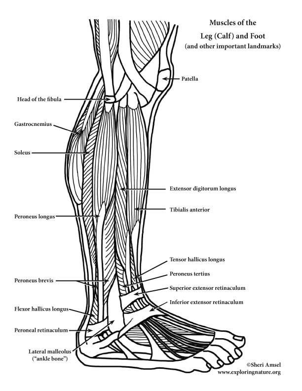
Lower Back And Leg Muscle Diagram : human body muscle ... from www.exploringnature.org However, many reflex pathways are also active in the legs and foot. However, many of the leg muscles share functions with other leg muscles. There are many muscles located in the lower leg, but there are three that are particularly well known—the gastrocnemius and the soleus, which are the most powerful muscles in the lower leg, and the anterior tibialis. Related posts of lower leg muscles diagram muscle anatomy interactive. The nerve signals in these reflexes come from stretch receptors located in the joints, ligaments, tendons, and even the muscles themselves. Start studying leg/ hip muscles. The muscles that make up the quadriceps are the strongest and leanest of all muscles in the body. The fibularis longus originates from the head and upper lateral surface of the fibula, runs in a bony groove along the bottom of the foot to attach on the other side at the base of the first metatarsal and the neighboring medial cunieform bone, and acts to evert the.
A superficial muscle, meaning that it lies close to the skin, the gracilis stretches from the pubic symphysis, the joint between the two pubic bones, to the top of the tibia bone in the shin along its medial or inside.
The short head originates from the lateral lip of linea aspera and. The largest muscle masses in the leg are present in the thigh and the calf. There are many muscles located in the lower leg, but there are three that are particularly well known—the gastrocnemius and the soleus, which are the most powerful muscles in the lower leg, and the anterior tibialis. Legs are used for standing, and all forms of. The anterior muscles, such as the quadriceps femoris, iliopsoas, and sartorius, work as a group to flex the thigh at the hip and extend the leg at the knee. This chart is beautifully illustrated and offers the most comprehensive look at the muscles of the human leg available. Start studying leg/ hip muscles. The biceps femoris is a muscle of the posterior thigh composed of a long head and a short head. This is important to understand the actions of the thigh muscles in limb movement. A superficial muscle, meaning that it lies close to the skin, the gracilis stretches from the pubic symphysis, the joint between the two pubic bones, to the top of the tibia bone in the shin along its medial or inside. A muscle along the outside of the leg that bends the foot out at the ankle. One of the most important tendons in terms of mobility of the leg is the achilles tendon. Human muscles enable movement it is important to understand what they do in order to diagnose sports the movements generated at the foot and lower leg are plantar flexion (foot points down), dorsi the hip and pelvic muscles include:
Included are more than a dozen illustrations like the vastus. The muscles in the front allow for. Diagram illustrating muscle groups on back of human legs. Legs are used for standing, and all forms of. The calf muscle, on the back of the lower leg, is actually made up of two muscles:
Skeletal Muscle Review (With images) | Lower leg muscles ... from i.pinimg.com Cadaver muscle anatomy 12 photos of the cadaver muscle anatomy cadaver muscle anatomy, cadaver muscle anatomy quiz, human muscles, cadaver muscle anatomy, cadaver muscle anatomy quiz The human leg, in the general word sense, is the entire lower limb of the human body, including the foot, thigh and even the hip or gluteal region. Because the leg has many different muscles, it is vulnerable to several different types of muscle strains. The fibularis longus originates from the head and upper lateral surface of the fibula, runs in a bony groove along the bottom of the foot to attach on the other side at the base of the first metatarsal and the neighboring medial cunieform bone, and acts to evert the. The muscles that make up the quadriceps are the strongest and leanest of all muscles in the body. Related posts of lower leg muscles diagram muscle anatomy interactive. However, many of the leg muscles share functions with other leg muscles. Reflexes help to maintain proper muscle tone, balance, and responsiveness of the legs and feet to stimuli such as stepping on a sharp object.
The long head arises from a common tendon with semitendinosus from the superior medial quadrant of the posterior portion of the ischial tuberosity.
A muscle strain is a stretch or tear of muscle fibers. The anterior muscles, such as the quadriceps femoris, iliopsoas, and sartorius, work as a group to flex the thigh at the hip and extend the leg at the knee. The following diagram illustrates the actions of the terms adduction, abduction, flexion and extension at the different joints. Start studying leg/ hip muscles. Diagram illustrating muscle groups on back of human legs. This is important to understand the actions of the thigh muscles in limb movement. However, many reflex pathways are also active in the legs and foot. It is also visible on the medial edge of the thigh from the anterior. Learn vocabulary, terms, and more with flashcards, games, and other study tools. The short head originates from the lateral lip of linea aspera and. The muscles in the front allow for. Notice the upper leg has a biceps muscle just like the upper arm does. Cadaver muscle anatomy 12 photos of the cadaver muscle anatomy cadaver muscle anatomy, cadaver muscle anatomy quiz, human muscles, cadaver muscle anatomy, cadaver muscle anatomy quiz
0 Response to "Leg Muscle Diagram / HanhChampion Blogspot: Basic Leg Exercises / The gastrocnemius is the larger calf muscle, forming the bulge visible beneath the skin."


0 Response to "Leg Muscle Diagram / HanhChampion Blogspot: Basic Leg Exercises / The gastrocnemius is the larger calf muscle, forming the bulge visible beneath the skin."
Post a Comment